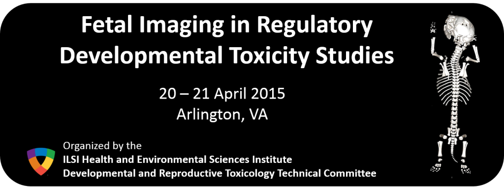
Fetal Imaging in Regulatory Developmental Toxicity Studies
- Start Date/Time :
- End Date/Time :
- Location : Arlington, Virginia, USA
Abstract
Developing and using specific imaging technologies (e.g., micro-CT, MRI) allows for alternative methods for evaluating structural birth defects in animal models. As the technology develops increased capabilities and speed, this technique could eventually become a high-throughput method and allow for sharing of images between laboratories and regulatory agencies. In practice, it will eliminate the use of large quantities of processing chemicals and wet-specimen archives. At this time, the technical methods, the alterations observed, and their interpretation have not yet gained general regulatory acceptance. In order to facilitate acceptance, a set of criteria will be developed that would help organizations validate their individual imaging systems and provide the basis for acceptance of these technologies by the regulatory community.
This workshop aims to:
- Introduce and discuss image capture technology relative to existing fetal evaluation methodology;
- Understand the regulatory community’s perspective and path for acceptance of results using imaging technology;
- Discuss minimal acceptable criteria for imaging to comply with Good Laboratory Practices and Computer Validations requirements; and
- Develop criteria to demonstrate concordance between new and existing examination methods and between testing results.
The final workshop program is available here.
Workshop Sessions
Monday, 20 April
Session 1: Background and Introduction
- Introduction to workshop & current paradigm for fetal evaluations in regulatory developmental toxicology studies, Howard Solomon, GlaxoSmithKline
- Introduction to imaging technology, Antong Chen, Merck
Session 2: Using micro-CT for Fetal Skeletal Evaluations
- Micro-CT methodology, Hasan Alsaid, GlaxoSmithKline
- Using micro-CT for fetal evaluations - an alternative to alizarin red staining when evaluating fetal skeletons, Sandra Wood, Merck
Session 3: "Concordance" Criteria - How Do We Define Scientific Agreement?
- Perspectives on concordance between the micro-CT as an alternative to tradtional staining methods, Julian French, Morphology Consulting, Inc.
- Comments on use of micro-CT technology in DART, Xiaoyou Ying, Sanofi
Tuesday, 21 April
Session 4: General Principles of Validation
- Regulatory perspectives - GLP (21 CFR Part 58) acceptance criteria, Zhou Chen, US FDA
- Regulatory perspectives - 21 CFR Part 11 Compliance, Johnathan Helfgott, Stage 2 Innovations
- GLP validation of technologies, software and procedures, Lelia Scott, Charles River Laboratories, Regulatory Affairs Compliance
Session 5: Summary & Discussion of Future Issues
- Longitudinal study to follow-up a developmental abnormality, Luc De Schaepdrijver, Janssen R&D
- Automated Skeletal Analysis of Rabbit Fetus Micro-CT images in DART studies, Belma Dogdas, Merck
- 3D Imaging and Automated Analysis of Mouse Embyro Phenotypes, Mark Henkleman, The Hospital for Sick Children and University of Toronto
- High throughput imaging modalities for phenotyping structural birth defects in mice, Cecilia Lo, University of Pittsburgh
Please contact Dr. Connie Chen (cchen@hesiglobal.org) for additional information.
